- Description
Specifications
MA:
200ma
Type:
Stationary
2. Rotary anode X-ray tube unit tangential annular tubes,
3. Single-phase full-wave rectification high-voltage generator
4. Power volage(V) photograph kilovolt(kV),infinitely variable control and electric mechanism
5. Be equipped with the manostat for the filament of X-ray tube and space charge complementor
6. Photographic volume, kV, mA and s, subsection, grading and interlock protection
7. Adopt the digital circuit timer. Grading according to R10 pririty coefficient, which is exact in time control.
8. High-voltage primary uses the zero controlled circuit of silicon-controlled rectifier of large power
9. Photographic bed of the 200mA Medical X-ray Equipment can move in length and breadth.
10 The photographic bed, upright and vibrating ray-filter of the 200mA Medical X-ray Equipment are in a whole without top and bottom track.
Specifications,
| Power supply | Adjustable range | With power supply 380V 340V—420V | |||
| With power supply 220V 195V—250V | |||||
| Fluoroscopy | Adjustable range of tube voltage | 50~95kV (Can adjust continuously) | |||
| Adjustable range of tube current | 0.5~5mA (Can adjust continuously) | ||||
| Adjustable range of tube voltage | 50~100kV (Can adjust continuously) | ||||
| Adjustable range of tube current | Small focus: 50mA | ||||
| Radiography | Big focus: 50, 100, 150 and 200mA | ||||
| Tube current of spot-film | 150mA (fix) | ||||
| Adjustable range of time | 0.04~6.3s 23 grades together | ||||
| High-voltage | Max. DC output voltage | 100kV | |||
| Generator unit | Max. DC output current | 200mA at instant 5mA at running | |||
| X-ray tube | Model | XD51-20.40/100 | |||
| Focus | 1mm×1mm 2mm×2mm | ||||
| Rotary angle | +90°~0°~-5° | ||||
| Move range of gastrointestinal spot-film device (fluorescent screen) |
Pressure direction: 300mm | ||||
| Horizontal direction: the center of diagnostic bed moves left (right) |
±80mm | ||||
| Vertical direction: 400mm | |||||
| Diagnostic bed | Ray-filter for fluoroscopy | Manual single blade | |||
| Distance between focus and bed face: | |||||
| At fluoroscopy and gastrointestinal radiography: 400mm(fix) | |||||
| Ray-filter tube (device) for radiography (fix X-ray field) |
Big (rectangle) | 14”×17” | Distance between focus and film=1m |
||
| Middle (rectangle) | 10”×12” | ||||
| Small (circle) | φ5” | ||||
The standard Configuration is without Chest Bucky Stand, It is optional
Chest Bucky stand Information
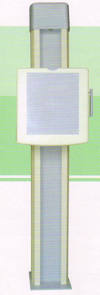
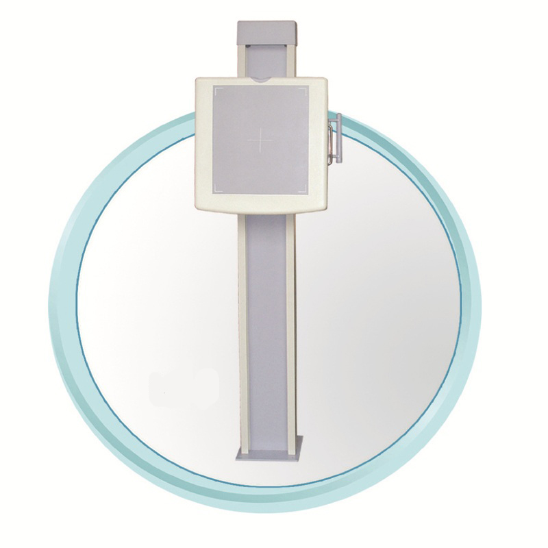
Consists of:
This device consists of grids, cassette drawers, columns, sliding frames and other parts.
1 column
2 sliding frame
3 cassette drawer
4 brake handle
5 groove pulley
6 weight screws
7 floor
8 suspension wire rope
Installation instructions
2.1 Safety Tips:
Equipment, installation and operation of the relevant units, the installation must comply with.
This photo frame is a monolithic chip packaging, the product will balance the heavy hammer before the factory bolt for easy handling, the bolt removed before installation.
2.2 Room conditions:
Environmental conditions: 0 ~ 40 ℃
Relative humidity: ≤ 75%
Atmospheric pressure: 700kpa ~ 1060kpa
Room height: 2700mm above the door width of the receiving equipment room door size of 1200mm width dimension of not less than 900mm
Room construction: room walls, doors and windows of the X-ray protection should comply with relevant regulations, in addition to the environment must fully consider the room temperature, humidity, clean degree, in order to protect the life and appearance of products. Room may not be used within the flammable gas or liquid.
2.3-ray bucky stand in the engine room to determine the installation position must meet the following conditions:
2.3.1-ray film holder in the engine room installation location, you must maintain the holder panel centerline alignment radiography X-ray Center.
2.3.2 Radiography rack-mounted film cassette to the X-ray tube to keep the focus away from the nearest point of 1000mm, the farthest point of ≥ 1500mm.
2.3.3 The ray film photography bed frame and supporting the use of only the X-ray tube head rotated 90 °, to open limit beam splitter lighting to make the limited cross-line alignment beam splitter light chest film frame panel can be centerline, while pulling Open tape, you can easily measure the distance between the focus away from the film.
2.4 that the selected election radiography and X-ray tube frame
To determine the best location between the base position, the water
Mud-based area of not less than 500 × 500mm.
2.4.1 Setting cement base, cement-based subject to the level of
Scale calibration level degrees.
2.4.2.Drill two M8 × 94 I-type expansion bolt hole, and screw in the expansion bolt buried.
Pacakge Information of this Chest stand,
1 Wooden Package, 208*60*46cm, Gross Weight: 127Kg, Net Weight: 100Kg
More detail information about the verticle chest stand, please use our user manual.
/u_file/1803/file/5301a945fe.pdf
Shipping Information:
G.W: 695kg
Packing Size: 2.45m,1.15m,1.12m
Unit: Piece
Special: No
Name:
Email:
Whatsapp/Tel:
Message:
 USD
USD EUR
EUR GBP
GBP CFA
CFA
